SportHorses.vet is an independent equine veterinary service based in Bocholt (3950), Belgium.
Specialized in equine orthopedics, sport horse medicine and performance-oriented veterinary care across Europe.


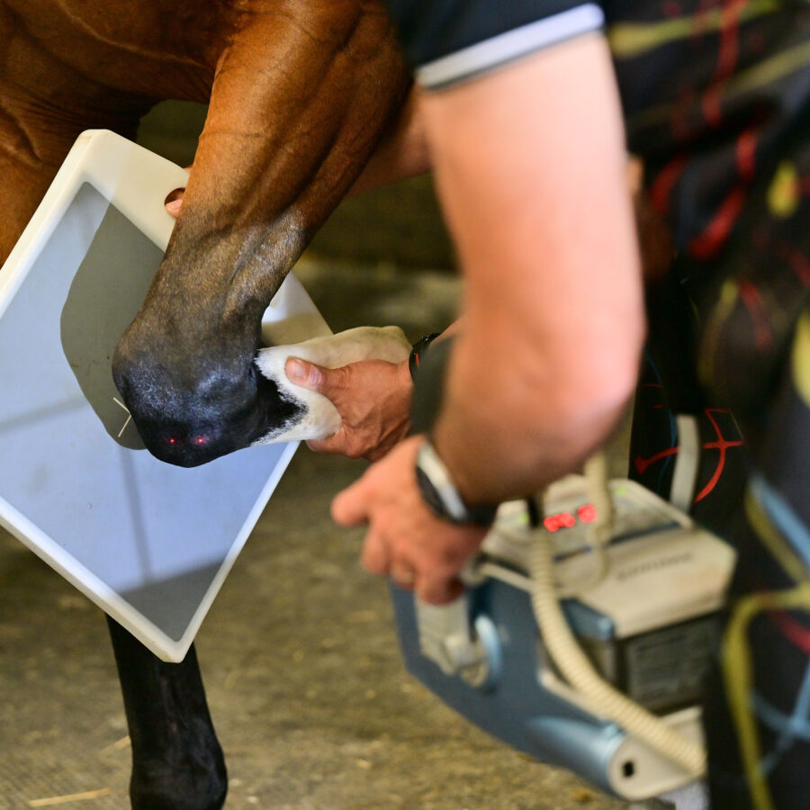
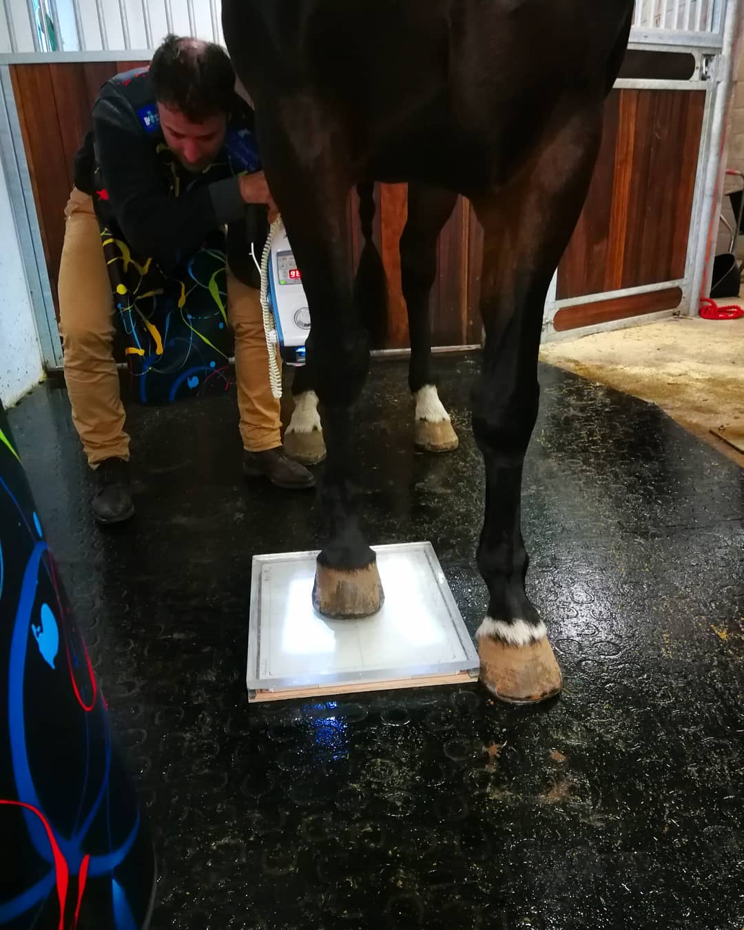
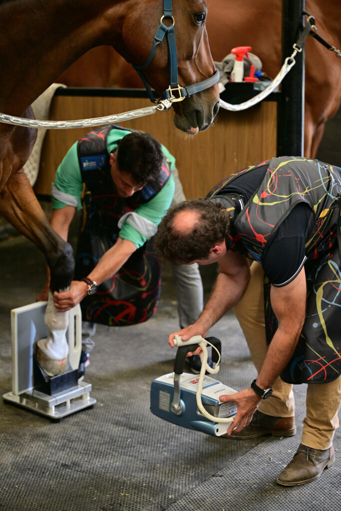
Radiography (X-ray imaging) is one of the most valuable and widely used diagnostic tools in equine orthopedics. Through a fast, non-invasive technique, radiographs allow us to visualize the internal structures of the horse—bones, joints, and specific anatomical details—making it possible to detect orthopedic problems early and improve treatment outcomes in sport horses.
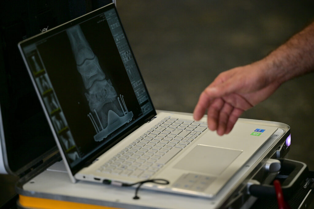
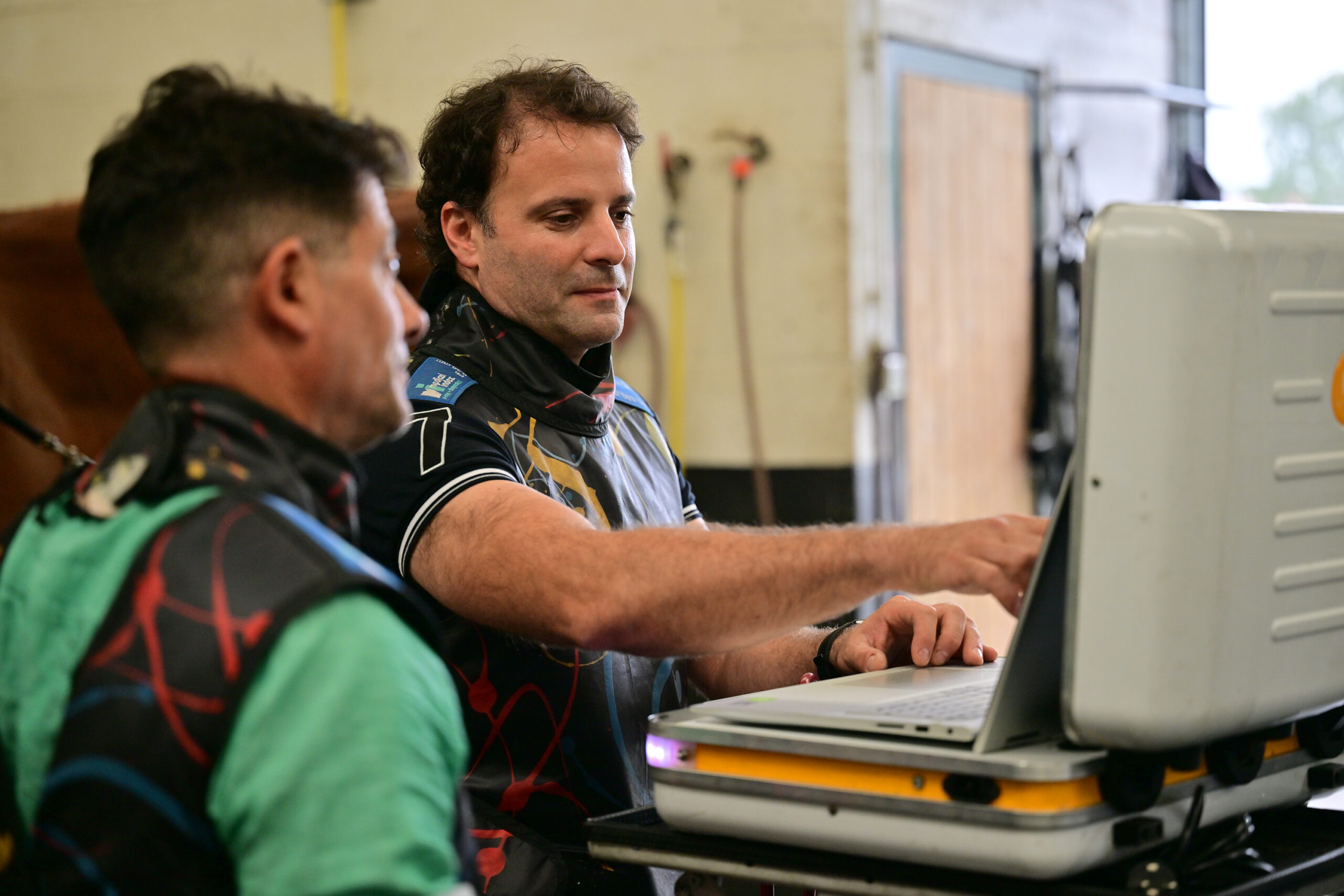
As an equine orthopedic veterinarian (PhD) based in Belgium, I use high-frequency digital radiography to obtain immediate, high-resolution images essential for accurate diagnosis, lameness evaluation, performance monitoring, and pre-purchase examinations across Europe.
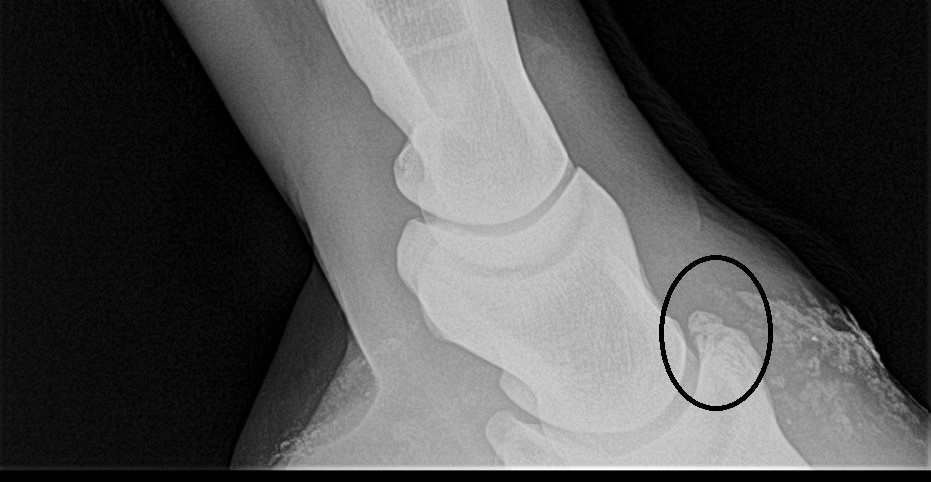
Radiographs are fundamental in diagnosing:
Early identification of these problems is crucial to prevent long-term damage and maintain the performance, longevity, and safety of sport horses.
Our advanced high-frequency digital radiography systems provide:
This technology ensures fast, precise and reliable diagnostic imaging—allowing us to make informed decisions for treatment, rehabilitation, and competition management.
Equine Orthopedic Veterinarian
Bocholt 3950, Belgium
2023 © All rights reserved.
SportHorses.vet is an independent equine veterinary service based in Bocholt (3950), Belgium.
Specialized in equine orthopedics, sport horse medicine and performance-oriented veterinary care across Europe.