


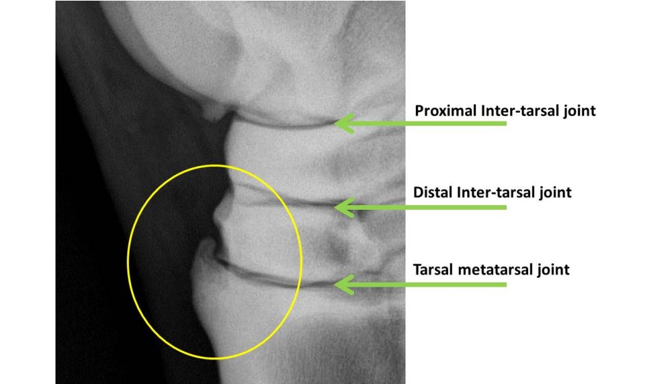
Problems distal to the hock, often referred to as “bone spasm”, is the most common cause of clinical hock-associated lameness in horses. Distal to the hock, osteoarthritis and periostitis of the distal intertarsal, tarsometatarsal, and occasionally proximal intertarsal joints occur.
The Hock consists of 4 joints: the tibiotarsal joint, the proximal intertarsal joint, the distal intertarsal joint, and the tarsometatarsal joint. During movement, the ankle joint performs approximately 98% of the hock motion. The other joints that make up the hock only have 2% movement.
Although the distal hock joints are not essential for normal locomotion in the horse, they are prone to instability. Chronic joint instability results in the development of joint inflammation (arthritis). Repeated compression and rotation of the tarsal bones and excessive tension at the attachment of the major dorsal ligaments have been implicated as causes of distal hock arthritis.
Factors that may affect the development of these pathologies distal to the hock include age, weight, breed, job description, frequency of work, intensity of work, and conformation of the horse.
The following are conformational abnormalities that may increase the chances of the horse developing distal hock arthritis:
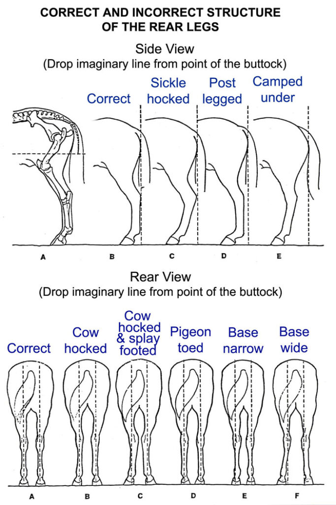
_Straight pelvic limbs: abnormal hock angle places foot too far back
_sickle hocks: This abnormal angle of the hock places the foot too far forward
_cow hocks: This defect results from the faster growth of the medial section of the tarsal bones than the lateral part. This structure creates tension inside the hocks
Clinical signs of arthritis/osteoarthritis distal to the hock joints usually show a gradual onset of lower limb lameness. The lameness is more evident during trotting and can be characterized by a hypermetric flight pattern of the hind limbs. Horses normally pull the pelvic limbs under their body and “stab” them outward when the foot strikes the ground surface. Although lameness is frequently bilateral, horse lameness favors the most affected limb during exercise. A “hip walk” (pelvic excursion) is often apparent, particularly when the horse trots with the affected limb inside a circle. The lameness may worsen after a period of rest. Affected horses usually show stiffness when they start to exercise, but can often “exercise.”
In chronic cases, a firm enlargement within the hock may become visible; the swelling represents excess bone associated with the distal tarsal joints. Horses with moderate to severe hock osteoarthrosis will generally exhibit a positive result. Churchill’s Hock test, a procedure performed during the evaluation of passive lameness. A positive response to this test is manifested by abduction of the lower limbs. Pelvic limb flexion (“Spavin test”) during active lameness assessment is an accurate and widely used detector. A positive response results in increased lameness and hypermetry after 60 to 90 seconds of lower limb flexion before jogging.
It is very common for horses to exhibit secondary symptoms as a result of favoring one or both hind limbs for an extended period of time. Common compensatory problems include:
_Ebaxial thoracolumbar (back) pain due to asymmetric lower extremity gait
_Suspensory desmitis of the proximal thoracic limb as a result of chronic overload of the thoracic limbs
_Greater trochanteric bursitis, as a result of abnormal gait of the pelvic limbs
Increased wear on the outside of the pelvic foot or shoe, in an attempt to relieve hock pain
Since these abnormalities are often secondary to distal osteoarthritis, successful treatment of hock will result in resolution of these problems.
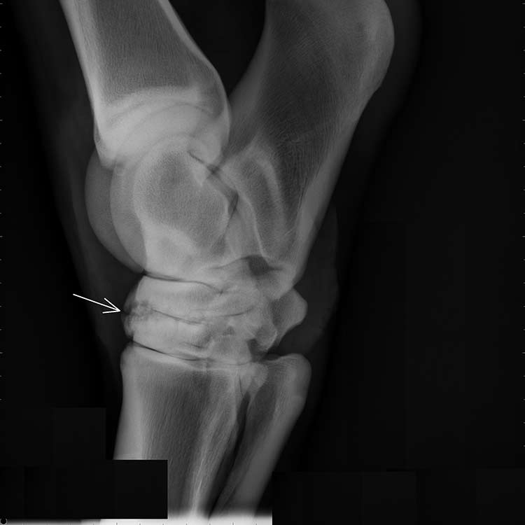
Diagnosis is primarily through clinical examination (demonstration of distal tarsal joint pain is diagnostic), lameness characteristics, response to Churchill hock test and hock flexion, and response to anesthesia. intraarticular. Radiographs are frequently used to assess the presence and severity of arthritis or osteoarthrosis from the hock to distal. However, it is important to note that joint inflammation alone (arthritis) is invisible on an X-ray (which provides only structural information). Nuclear scintigraphy (bone scan) may be a more accurate assessor of the presence of distal tarsal inflammation, as it provides functional information (ie, measures blood flow).
For the treatment of distal osteoarthrosis of the hock there are two basic forms of treatment:
_The first involves the reduction and possibly elimination of inflammation within the distal tarsal joints. This is achieved through the use of systemic and/or intra-articular anti-inflammatory therapy.
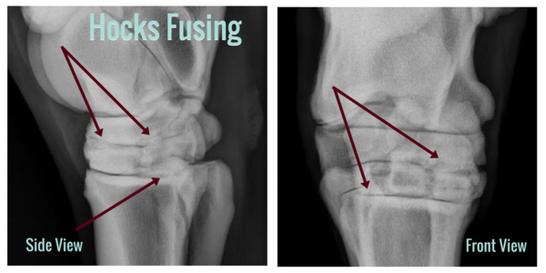
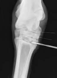
The other form of distal tarsal therapy involves fusion of the distal intertarsal and tarsometatarsal joints. This can be achieved surgically or through the use of a chemical agent that is infused into the distal tarsal joints to eliminate low mobility joints, also eliminating instability and therefore inflammation and pain.
If you would like more information on treatment options for your horse’s hock, please do not hesitate to contact Sporthorse.vet we will be happy to discuss this topic in more detail.
No Comments