


Injury to the ventral discs on occasions is accompanied by bone, ventrolateral or lateral proliferation (vertebral spondylosis); It is found mainly in the mid-thoracic area (mainly between T11 and T13) and also in the lumbar area.
Spondylosis is a rare cause of back pain in horses is around 3.5%
They can be found in asymptomatic horses, but they can also be responsible for acute pain or chronic stiffness of the back.
The causes of hindlimb ataxia and lumbosacral pain are often ambiguous, difficult to diagnose, and of uncertain etiology, which may arise from spontaneous musculoskeletal disease, neurologic dysfunction, or traumatic injury.
Much of the clinical signs associated with equine spinal disc problems are neurological.
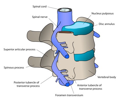
Lumbosacral joint is the most mobile intervertebral joint between the skull thoracic vertebrae and pelvis in the horse. This mobility makes the lumbosacral joint more susceptible to injury than others intervertebral joints.
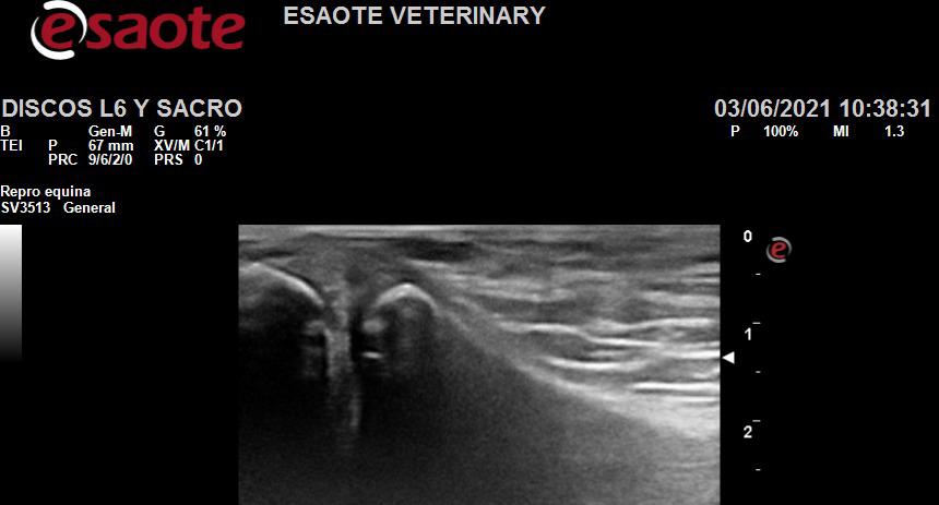
Spinal discs in horses in L5-L6
With ultrasound, the alterations are revealed as changes in the
echogenicity of the lumbosacral disc, multifocal areas of mineralization within the lumbosacral disc.
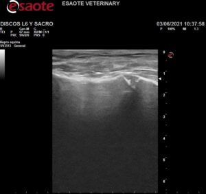
Mild bone proliferation alterations affecting the L6-S1 intertransverse joints such as asymmetry of the width of the sacroiliac joints and osteophytosis present.
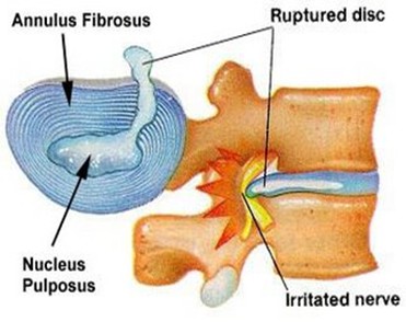
A diffuse loss of echogenicity, fiber breakage, fiber degeneration, disc fissure, disc cavitation, scar tissue, spots that may be indicative of previous lumbosacral trauma or change secondary to mineralization of the intervertebral disc.
The low presentation of clinical disc disease in the horse is attributed to the absence or underdeveloped nucleus pulposus, and relatively thin width of intervertebral discs in equines.
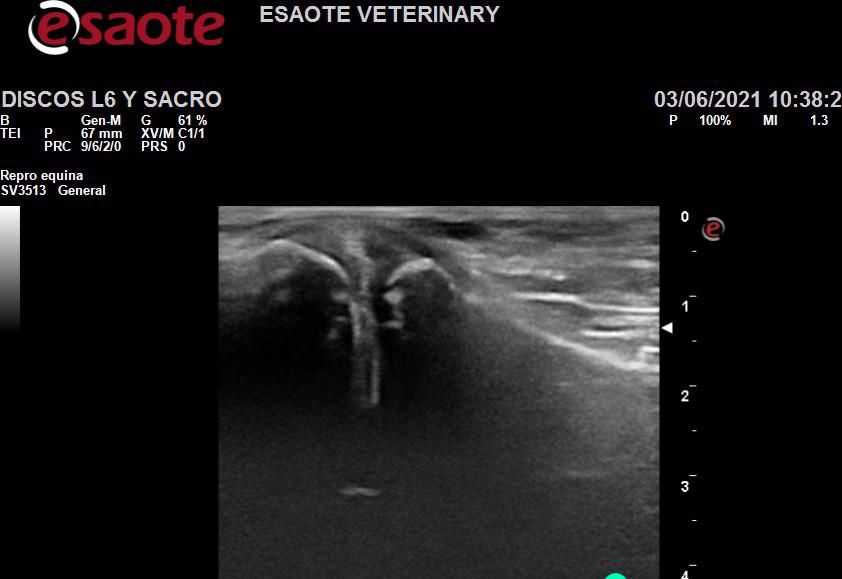
Spinal discs in horses L6-S1
The intervertebral discs have a central fibrocartilaginous area that becomes more fibrous peripherally and is relatively thin,
making horses less susceptible to disc prolapse.
It is necessary to mention the importance of the sacrocaudalis dorsal and multiple fi dus muscles that help further stabilize and support the lumbosacral area subjected to dorsoventral movement.
Kinematic evaluation of the lumbosacral space reveals a wide range of dorso-ventral mobility of the joint that goes 9 +/- 32 ° of L5-S1 and is attributed to increased thickness and lower height of the lumbosacral intervertebral discs.
Lumbosacral instability can have it predisposed this horse to injury and disc protrusion which predisposes the horse to cauda equina compression or L6-S1 instability.
Spinal discs in horses
For the ultrasound diagnosis of the intervertebral lumbosacral in disc disease requires careful evaluation and knowledge of normal age-related changes in horses in addition to changes in echogenicity, shape, positioning of the intervertebral disc
and correlation of clinical signs. Also, horses diagnosed with lumbosacral or sacroiliac pain may warrant more investigation and follow-up using nuclear scintigraphy to correlate degenerative changes with inflammatory changes or remodeling associated with the lumbosacral area.
No Comments